Effect of Photoperiod and Melatonin Administration on the Midgut of the Nocturnal Insect Spodoptera litura
Effect of photoperiod on developing individuals in Spodoptera litura
DOI:
https://doi.org/10.5530/fra.2025.1.3Keywords:
Melatonin, Histology, Photoperiod, Midgut physiology, InsectsAbstract
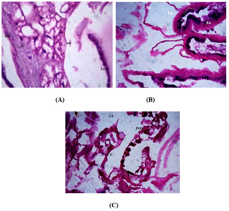
Background: Melatonin is known to be powerful antioxidant in animals. However, few studies have been conducted to verify the protective effect of melatonin in insects. This study's objective was to interpret the changes in photoperiod with response to melatonin and luzindole in midgut physiology of S. litura. Materials and Methods: Third instar S. litura larvae (N=180) were maintained with different types of treatment follows, i) Control (phosphate buffer saline; pH 7.5); ii) Melatonin (4.3 × 10⁻⁵ M/100 mL diet) and iii) Luzindole (is 250 mM/100 mL diet); under Photoperiods (24L: 0D; 12L: 12 D and 0L: 24 D) in Molecular entomology Laboratoy, Periyar University, Salem during March 2016-July 2017. The developmental parameters and Reproductive attributes of S. litura were statistically analyzed. Treated S. litura midgut tissues were stained with hematoxylin eosin, and photographed under Phase Contrast microscope. Results: Results indicate that 12L: 12D showed the cellular injury in the lumen and columnar epithelial cells, whereas in 0L: 24D, intercellular spaces expanded along with the columnar cells were detected in melatonin and luzindole treatments. When larvae treated with luzindole, histological changes such as cytoplasm vacuolization, breakdown of the brush border membrane, vesicle formation were examined. The control larvae of S. litura kept under 0L: 24D and 24L:0D which displayed a layer of columnar and epithelial cells that was well maintained. Conclusion: This work has demonstrated the distinct impacts of luzindole and photoperiod on the histology and developmental fitness of the nocturnal insect S. litura.
Downloads
Metrics